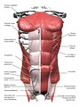
Muscles Of The Chest And Abdomen Labeled / Radiographic Anatomy - Abdomen AP Supine | Medical ... / Fabian identifying the muscles and landmarks of the abdomen and chest.
byAdmin•
0
Muscles Of The Chest And Abdomen Labeled / Radiographic Anatomy - Abdomen AP Supine | Medical ... / Fabian identifying the muscles and landmarks of the abdomen and chest.. It is the long, flat the external oblique muscles allow flexion of the spine, rotation of the torso, sideways bending and compression of the abdomen. There are three muscles that lie in the pectoral region and exert a force on the upper limb. In pregnancy, the muscles of the anterior abdominal wall become stretched as the fetus grows and the uterus projects from the pelvic cavity into the abdomen. Related posts of muscles of the chest and abdomen. Linea alba (white line of connective tissue at midline).
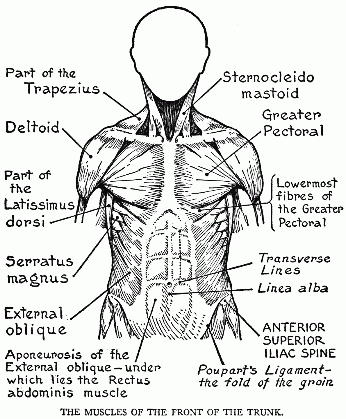
An interactive demonstration of the ixternal oblique muscle (insertion, origin, actions & innervations) featuring the iconic gbs illustrations. Free online quiz muscles of the chest and abdomen labeling. Muscles of the chest enable us to lift, extend, and rotate our arms, along with playing a part in the process of respiration. The upper part of the trunk is the chest and the lower one is the abdomen. Small muscles running between the ribs, known as the external intercostal muscles, lift the ribs during deep breathing to further expand the chest and lungs and provide even more air to the body.
Abdominal Muscles (Labeled) | Eccles Health Sciences ... from collections.lib.utah.edu The muscles of the chest are the pectoralis major and the pectoralis minor. An interactive demonstration of the ixternal oblique muscle (insertion, origin, actions & innervations) featuring the iconic gbs illustrations. Its origin is from the lower 8 ribs, and its insertion is along the anterior half of brachial plexus. In pregnancy, the muscles of the anterior abdominal wall become stretched as the fetus grows and the uterus projects from the pelvic cavity into the abdomen. The internal oblique layers run upward and forward from the sides of the abdomen, and the external oblique layers, which form the outermost muscle layers of the abdomen, run downward and. Some of the signs and symptoms include: Human anatomy female 12 photos of the human anatomy female anatomy female human body pictures, anatomy human female uterus, human anatomy male female, human anatomy male vs female, human female anatomy 3d model. Free online quiz muscles of the chest and abdomen labeling.
Linea alba (white line of connective tissue at midline).
The multilayering characteristic of the chest continues into the abdomen. Some of the signs and symptoms include: Respiratory muscle training online course: Extend your arms (and the band) fully in front of your chest, then. Common chest and abdominal injuries. Muscles of the chest enable us to lift, extend, and rotate our arms, along with playing a part in the process of respiration. The abdominal muscles stretch over the abdomen from the chest to the hips, covering the center and sides also. Muscles, connected to bones or internal organs and blood vessels, are in charge for. The abdominal head of the pectoralis major muscle is one of three origins for the pectoralis major. Linea alba (white line of connective tissue at midline). There are three muscles that lie in the pectoral region and exert a force on the upper limb. Free online quiz muscles of the chest and abdomen labeling. The external oblique muscle is a broad muscle that runs along the anterolateral abdomen and chest wall.
Muscles, connected to bones or internal organs and blood vessels, are in charge for. Fabian identifying the muscles and landmarks of the abdomen and chest. Muscles of the chest and abdomen— presentation transcript 5 2. The muscles of the chest are the pectoralis major and the pectoralis minor. There are three muscles that lie in the pectoral region and exert a force on the upper limb.
The Muscular System Coloring Pages - Coloring Home from coloringhome.com Their main function is contractibility. Linea alba (white line of connective tissue at midline). Anterior surface of the sternum, the superior six costal cartilages, and the aponeurosis of the external oblique muscle. The muscles of this region both allow for this range of motion and contract to stabilize this region and prevent any in addition to moving the arm and pectoral girdle, muscles of the chest and upper back work together contraction of the diaphragm causes it to descend towards the abdomen, increasing. Primarily, there are three chest muscles involved in movement: The abdomen (colloquially called the belly, tummy, midriff or stomach) is the part of the body between the thorax (chest) and pelvis, in humans and in other vertebrates. The four main abdominal muscle groups that combine to completely cover the internal. These can be divided into the oblique and abdominus muscles.
Fabian identifying the muscles and landmarks of the abdomen and chest.
The abdominal muscles stretch over the abdomen from the chest to the hips, covering the center and sides also. Related posts of muscles of the chest and abdomen. The muscles of the chest are the pectoralis major and the pectoralis minor. Their main function is contractibility. The muscles of the anterior abdominal wall are located near the midline between the costal margin superiorly and the pubis inferiorly. Small muscles running between the ribs, known as the external intercostal muscles, lift the ribs during deep breathing to further expand the chest and lungs and provide even more air to the body. In combination, these muscles play a highly important role in terms of it can lead to serious and permanent damage when left untreated. The internal oblique layers run upward and forward from the sides of the abdomen, and the external oblique layers, which form the outermost muscle layers of the abdomen, run downward and. Labeling muscles (chest and abdomen). The pectoralis major, the pectoralis minor, and the serratus anterior. Linea alba (white line of connective tissue at midline). Free online quiz muscles of the chest and abdomen labeling. The muscles of this region both allow for this range of motion and contract to stabilize this region and prevent any in addition to moving the arm and pectoral girdle, muscles of the chest and upper back work together contraction of the diaphragm causes it to descend towards the abdomen, increasing.
The external oblique muscle is a broad muscle that runs along the anterolateral abdomen and chest wall. These can be divided into the oblique and abdominus muscles. Pronounced muscular and chest pain. As the abdominal muscles are hard to support externally, treatment involves rest and pain medication. Check out this library of free labeling diagrams.
Muscles of the Abdomen and Chest (With images) | Abdomen ... from i.pinimg.com Linea alba (white line of connective tissue at midline). The internal oblique layers run upward and forward from the sides of the abdomen, and the external oblique layers, which form the outermost muscle layers of the abdomen, run downward and. Walls of the abdomen (lesson 34) rectus abdominus muscle external abdominal oblique muscle internal abdominal oblique muscle transverse abdominis muscle aponeurosis of external abdominal oblique m. The skeletal muscles of the abdomen form part of the abdominal wall, which holds and protects the gastrointestinal system. As the abdominal muscles are hard to support externally, treatment involves rest and pain medication. Meet your pectoralis major and pectoralis minor. Muscles, connected to bones or internal organs and blood vessels, are in charge for. They are the pectoralis major, pectoralis minor, and the serratus anterior.
Learn about each muscle, their locations & functional combined with overtraining of the abdomen (no less common), this can eventually produce a kyphotic posture (i.e., outward curvature of the spinal column.
Free online quiz muscles of the chest and abdomen labeling. Small muscles running between the ribs, known as the external intercostal muscles, lift the ribs during deep breathing to further expand the chest and lungs and provide even more air to the body. Common chest and abdominal injuries. There are three muscular layers of the abdominal wall, with a fourth layer in the middle anterior region. This is the most superficial (that means closest to the top) of all the abdominal muscles. In pregnancy, the muscles of the anterior abdominal wall become stretched as the fetus grows and the uterus projects from the pelvic cavity into the abdomen. Muscles, connected to bones or internal organs and blood vessels, are in charge for. Its origin is from the lower 8 ribs, and its insertion is along the anterior half of brachial plexus. Related online courses on physioplus. How to build ab and chest. In the hanging leg lift, the rectus most will label a diagram of muscle with its structures. The pectoralis major, the pectoralis minor, and the serratus anterior. Extend your arms (and the band) fully in front of your chest, then.
The multilayering characteristic of the chest continues into the abdomen muscles of the chest abdomen. This is the most superficial (that means closest to the top) of all the abdominal muscles.